Basics Of Flow Cytometry
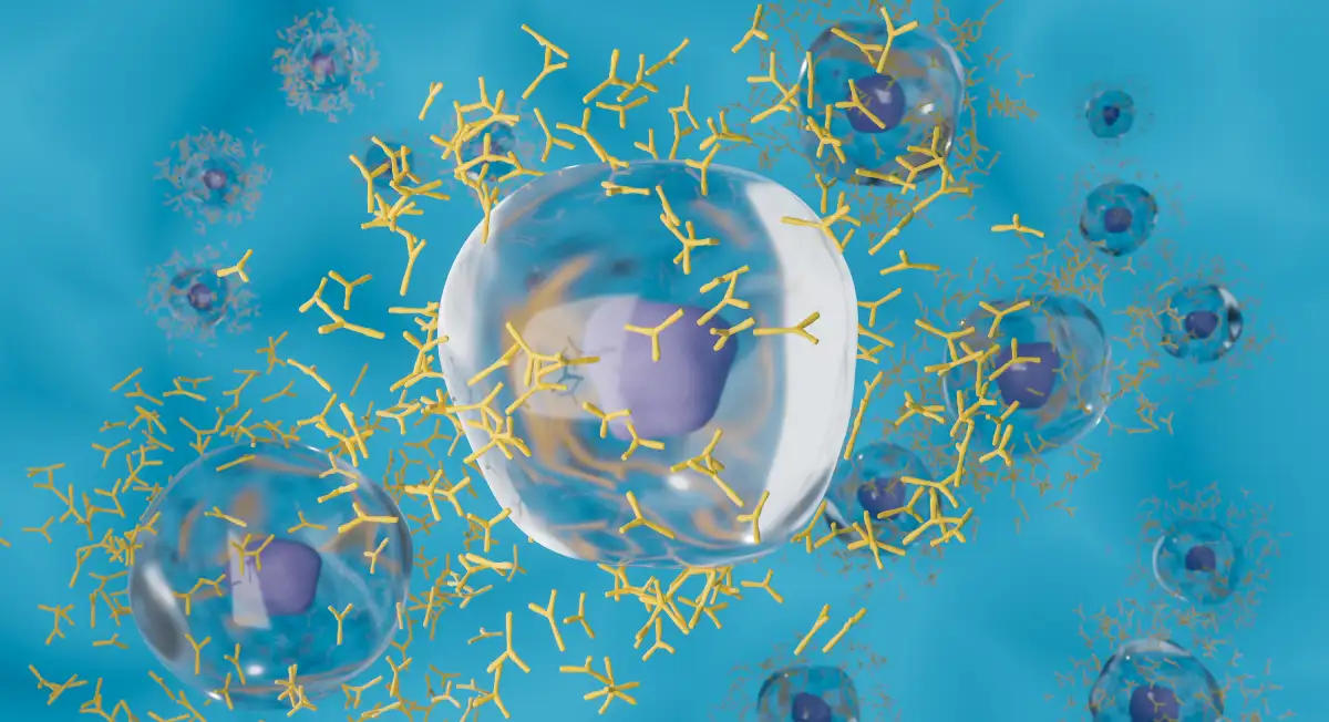
Now that we know the meaning of flow cytometry, this section will provide a concise overview of the working principles of flow cytometry, cytometry basics, and flow cytometry procedures. Flow cytometry works on the fundamental principle of light scattering, excitation, and emission when fluorescently tagged cells or cell components pass through a laser light. During the flow cytometry procedure, the technician prepares a monodispersed suspension of non-clustered cells tagged with fluorescent antibody labels (cells may be tagged externally or internally) and injects them into the flow cytometer. The cytometer consists of three components- fluidics, optics, and electronics. The fluidics is responsible for the flow of the sample; the optics component includes the laser source, optical filters, and wavelength detection system; and the electronic component is responsible for data acquisition and processing. The cells pass through the laser beam, and the instrument measures the scattered and fluorescent light. This method of flow cytometry using fluorescent labels is also known as fluorescence-activated cell sorting (FACS).
Principles Of Flow Cytometry
After a concise overview, let us understand the working principles of flow cytometry in detail. When the cell suspension flows through the fluidics system within the flow cytometer, the cells are focused hydrodynamically by the sheath fluid, and they pass in a single file through the narrow nozzle. The hydrodynamic focusing causes the cells to separate and align in a single file. This produces an extremely tiny fluid stream, almost equivalent to one cell passing at a time across the laser beam. Based on the flow cytometry principle, the detector in the optics component detects forward scattered (FS) and side scattered (SS) light, along with flow cytometry fluorescence emitted by the fluorescently tagged cells. The direction of light scattered by the cells is related to cell size and granularity; while FS correlates to cell size, SS correlates to cell granularity. Hence, expectedly, a sample containing large granulocytes will generate high FS and SS light, while monocytes having less granularity than granulocytes will produce high FS but low SS light. This is the underlying flow cytometry principle that enables cell sorting.
Applications Of Flow Cytometry
Flow cytometry is a crucial bioanalytical technique that finds diverse applications in medical and healthcare domains, research, and drug discovery. Notably, FACS is an indispensable research technique in hematopoiesis, oncology, immunotherapy, stem cell research, and clinical diagnostics.
Medical Applications
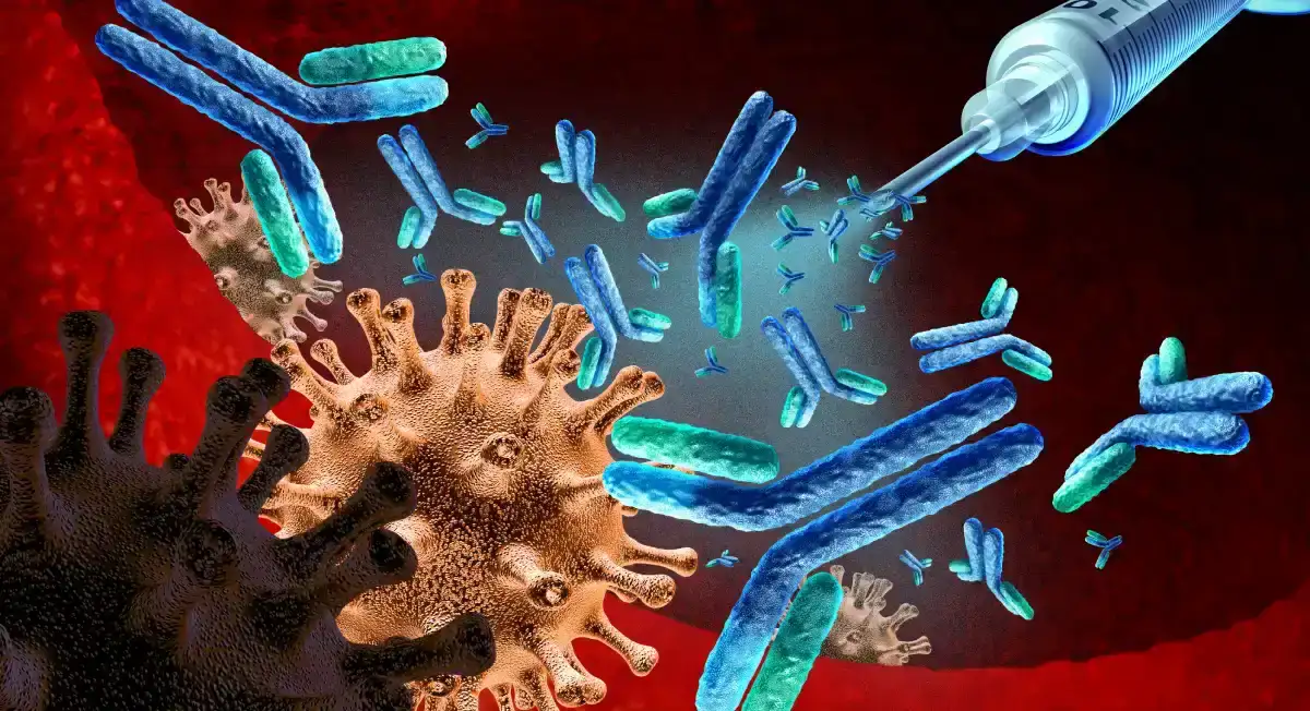
In the medical field, flow cytometry uses include diagnosis and developing treatment plans for various infectious diseases and cancer. The capacity of cell flow cytometry to characterize cells makes it a powerful tool for detecting specific morphological changes in cells and tissues associated with different diseases or their stages, thus diagnosing diseases or predetermining their recurrences. As a diagnostic tool, flow cytometry can detect blood disorders such as Paroxysmal nocturnal hemoglobinuria (PNH) and antithrombin deficiency, blood cancers such as leukemia and lymphoma, and immune disorders such as HIV and other inherited immune deficiencies. It does so by quickly detecting the increased lymphocyte population or any abnormal cell counts, characterizing its phenotype, and classifying the lineage of the cell population, thereby enabling the diagnosis of the disease.
Research Applications
In molecular science research, cytometry applications include the detection and characterization of protein expression, post-translational modifications of proteins, including cleaved and phosphorylated proteins, RNA, including mRNA transcripts and miRNA, cell health status, various stages of the cell cycle, and cell sorting in a heterogeneous sample.
Other Uses
Other flow cytometry applications include its use in environmental microbiology, where the technique helps to study the biology, ecology, and biogeochemistry of marine photosynthetic picoplankton and microbial populations in diverse matrices such as soil, water, and sediments. It is a versatile technique that helps improve our understanding of various ecological processes, assess the effects of humans on nature, and identify sources of pollution, thus assisting us in developing effective management strategies.
Types Of Flow Cytometry Assays
Since flow cytometry has diverse uses, various application-based flow cytometry assays have developed that allow apoptosis analysis, immunophenotyping, cytokine detection, cell cycle analysis, cell viability determinations, cell proliferation analysis, microbial analysis, and other functional arrays to detect cytokines, phagocytosis, immune function, etc.
Structural and morphological changes accompany apoptosis or programmed cell death. In the cytometry test for apoptosis analysis, the technique measures the characteristic changes in cell size, morphology, and DNA content to distinguish apoptotic cells from healthy cell populations.
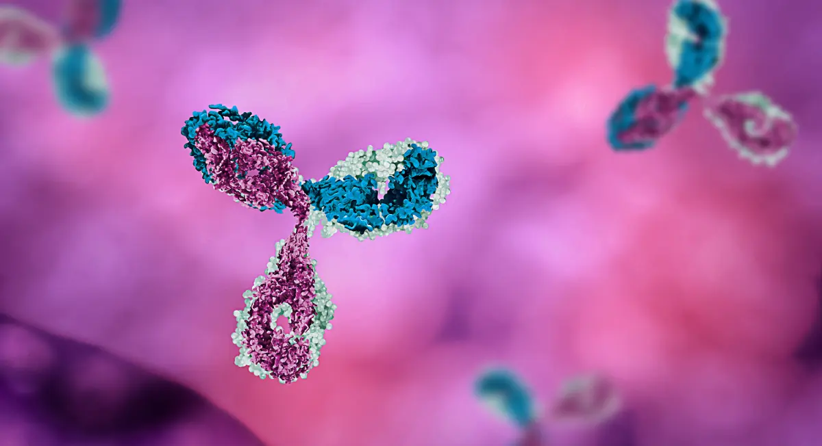
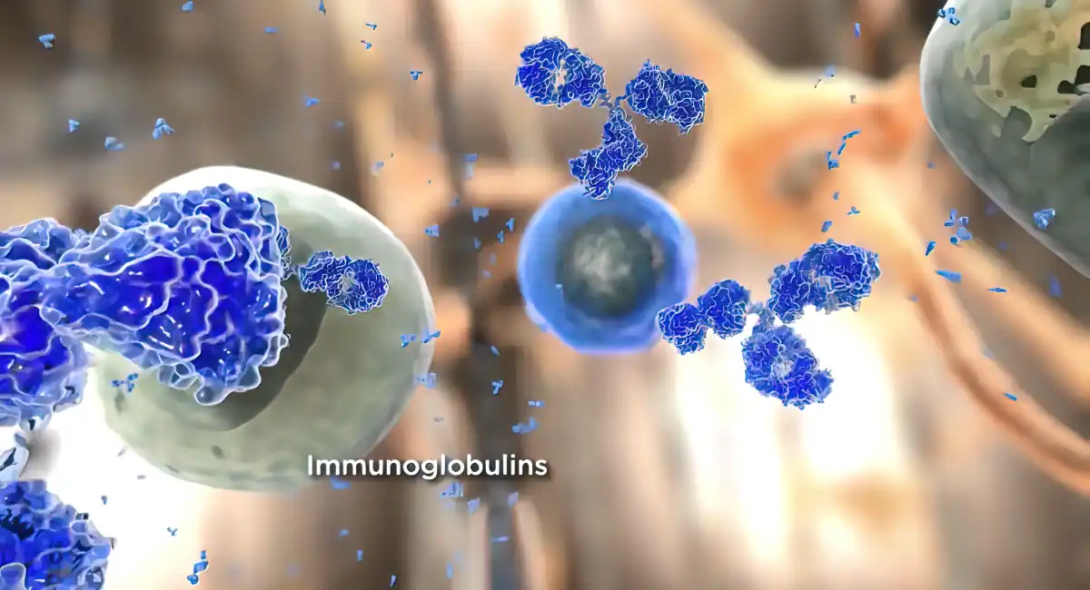
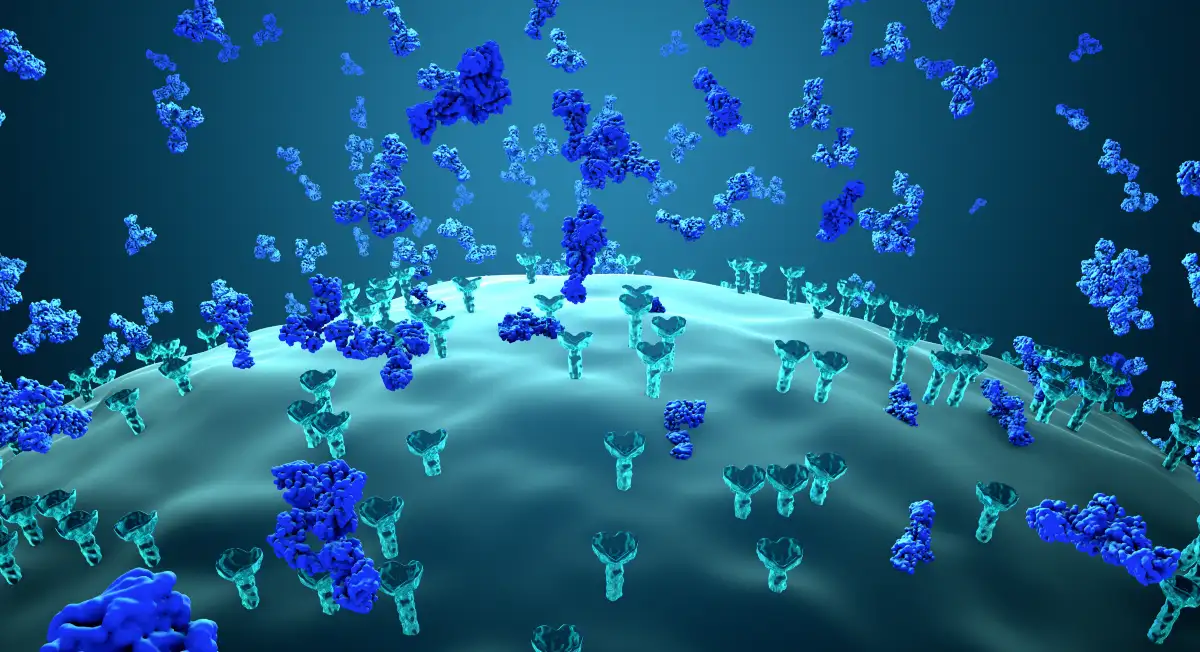
Immunophenotyping helps to understand the protein expression in cells by using cell or tissue samples. In immunophenotyping flow cytometry assay, the cell populations are identified and quantified based on protein-based biomarkers expressed on the cell surfaces. This is achieved by using specific antibodies directed against the cell surface proteins. Immunophenotyping assay helps to detect cancers such as leukemia.
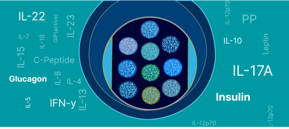
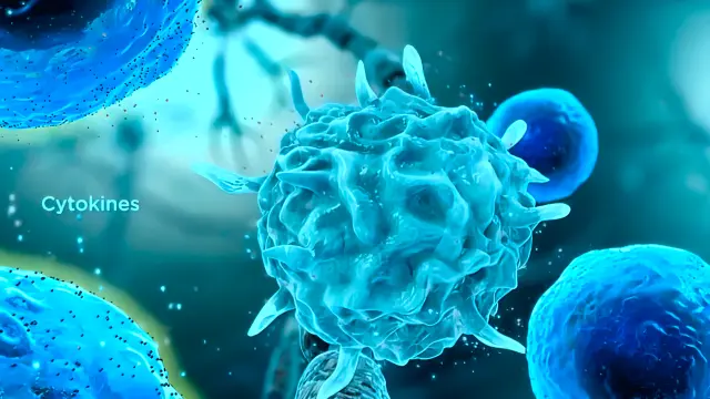
Similarly, flow cytometry assays are highly efficient in detecting intracellular cytokine secretion, even at the single-cell level. This technique employs fluorescently labeled monoclonal antibodies for intracellular cytokine staining. It measures multiple cytokines within a single cell and is thus capable of detecting complex phenotypes of cytokines.
Flow Cytometry Methodology
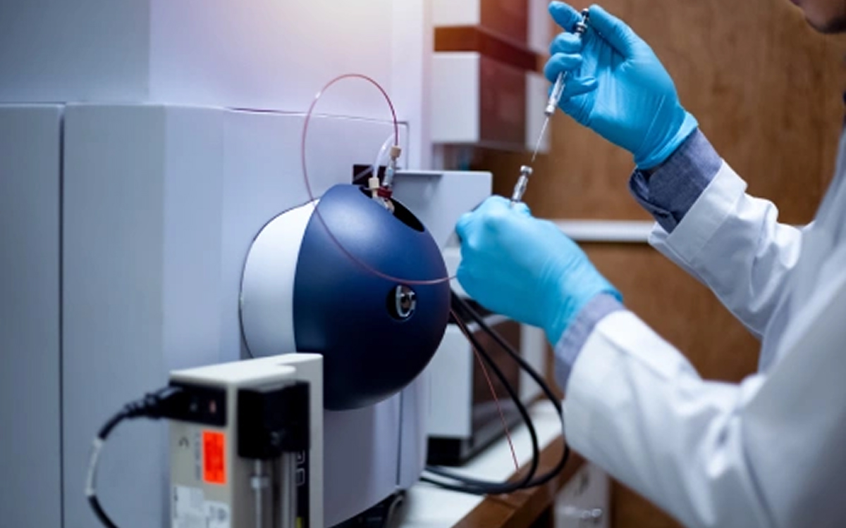
Sample Preparation
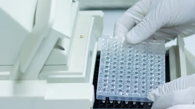
Flow cytometry analysis begins with sample preparation. Samples may include blood, bone marrow, organ tissues, or other biological specimens. Cells may be harvested through mechanical dissociation or enzymatic digestion of tissue followed by centrifugation and mechanical filtration to remove cell debris and cell clumps and avoid instrument clogs or poor data quality. Single-cell suspensions are prepared in cold staining buffer at a density of 106 – 107 cells/ml.
Data Acquisition
The single-cell suspension is introduced into the flow cytometer, and the cells in the sample pass through the laser beam after hydrodynamic focusing. The detectors placed in front and side of the laser beam measure the scattered light, while the fluorescence detector measures the fluorescence emitted by the stained cells. This process of collecting data is known as data acquisition. The flow cytometer measures the height of the total voltage and the area that equates to the fluorescence intensity per channel. Once the data is acquired, flow cytometry charts such as histograms and dot plots can be obtained.
Gating and Data Analysis
Having learned how to acquire data in flow cytometry, let us now see how to interpret flow cytometry data. A technique known as gating facilitates data analysis. Gating refers to the numerical or graphical boundary that denotes subpopulations of cells for further analysis. Gating is done by selecting the area on the dot plot or histogram generated during the experiment for analysis. Gating also helps to remove dead cells and doublets from flow cytometry analysis. Some gates used include quadrants, rectangular, elliptical, and polygons for dot plots. Since a gate indicates a subset of the total population, the total population and the percentage of the subpopulations in a heterogeneous sample can be estimated.
Advanced Techniques In Flow Cytometry
Multicolor Flow Cytometry
Leveraging the basics of flow cytometry, multicolor flow cytometry is a rapidly evolving advanced flow cytometry technique that uses multiple fluorescent markers to identify and characterize different subpopulations of cells. Since cells of different types have different antigens, they bind to different antibodies. This enables the distinction of various cell types based on the fluorescence they emit. Compared to flow cytometry, multicolor flow cytometry has higher efficiency with lower sample requirements.
Fluorescence-Activated Cell Sorting (FACS)
FACS cytometry is a technique that helps to separate different subpopulations of cells differentiated by flow cytometry based on their phenotypes. Thus, this cell sorting technique enables researchers to better understand each cell type in a heterogeneous cell sample. FACS cytometry is commonly used to isolate stem cell populations or lymphocytes from white blood cells.
Recent Technological Advances
Previously, the use of flow cytometry technique was limited owing to the limited number of lasers and detectors, which restricted the simultaneous use of multiple fluorescent markers. With technological advancements, several optical lasers and their corresponding detectors are available. This has paved the way for developing advanced flow cytometry techniques with multiplexing capabilities.
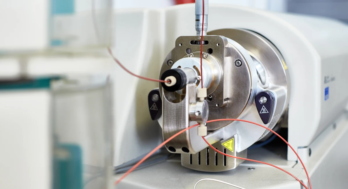
Key Components Of A Flow Cytometer
To get an overview of how a flow cytometer works, let us look at the flow cytometry machine and the components of flow cytometry. A flow cytometry machine has three main components- fluidics, optics, and electronics.
The fluidics helps to transport the sample from the sample tube to the flow cell. Once the sample flows past the flow cell, the cells in the sample are either sorted or discarded, depending on the application involved.
The optics include excitation sources, i.e., lasers, lenses, and filters. These components help to excite the sample and collect the scattered and fluorescent light for the detector system that produces a photocurrent corresponding to the event.
The electronics component is the brain of the flow cytometer. The photocurrent generated by the detector system is digitized and processed for subsequent analysis using software.
Interpreting Flow Cytometry Results
Once the experiment is done, data analysis is performed, and results are interpreted for flow cytometry explanation. By now, we know flow cytometry definition, so let us see how flow cytometry results are interpreted to understand what flow cytometry measures.
Normal vs. Abnormal Patterns
Flow cytometry helps to sort cells, along with phenotyping and characterization of different subpopulations of cells. This allows the technique to identify deviations from the normal antigen expression patterns in cells compared to healthy cells. A healthy cell will exhibit an antigen expression pattern according to its type and maturity. However, an abnormal cell displays aberrant antigen patterns that could indicate the presence of a disease. When these aberrant antigen patterns match specific disease biomarkers, the associated disease can be easily identified.
Common Conditions Diagnosed
Abnormal antigen patterns in flow cytometry can help to identify conditions such as acute lymphoblastic leukemia, acute myeloid leukemia, chronic lymphocytic leukemia, multiple myeloma, and non-Hodgkin lymphomas (both B-cell and T-cell).
Advantages And Limitations Of Flow Cytometry
Flow cytometry is a powerful technique that enables the phenotyping and characterization of cells at a single-cell level. The advantages of flow cytometry include its high speed, accuracy, capacity for single-cell analysis, identification of a small population of cells within a heterogeneous sample, quantification based on fluorescence intensities, and sorting of cells. Furthermore, advanced modalities like multicolor flow enable simultaneous identification and characterization of different subpopulations of cells. With advancements in technology and the integration of AI in flow cytometry, the advantages of flow cytometry will increase manifold. Despite these advantages, there are a few limitations. Flow cytometry limitations include the high cost of the sophisticated instrument, the need for specialists to manage the instrument and analyze complex data produced, and the need for single-cell flow for analysis. In addition, since the instrument is complex, it is prone to error. Thus, it needs proper cleaning and calibration before each use.
Future Trends In Flow Cytometry
Flow cytometry is a technique that finds immense application in medical and molecular science research. It is a powerful technology that allows comprehensive and multiplexed analysis of single cells. With rapid technological advancements, flow cytometry will play a transformational role in healthcare, including developing personalized medicine. The current trends in cytometry indicate that the future of flow cytometry will be guided by technological advancements such as the development of high-throughput systems and the integration of AI technology in flow cytometry, leading to the development of next-generation techniques. AI can improve the accuracy and efficiency of flow cytometry-based diagnosis in clinical settings and assist in data management.
Conclusion
This section provides a brief cytometry conclusion. In summary, flow cytometry service is powerful, having numerous applications ranging from phenotyping to determining cell health and viability. It is used in domains such as immunology, hematology, oncology, and microbiology and thus is an indispensable technology to clinicians and researchers. It operates on the fundamental principle of light scattering, excitation, and fluorescence emission by cell suspension when cells pass through a laser light. It is highly versatile, rapid, and capable of measuring multiple parameters of millions of cells with high accuracy at a single-cell level.