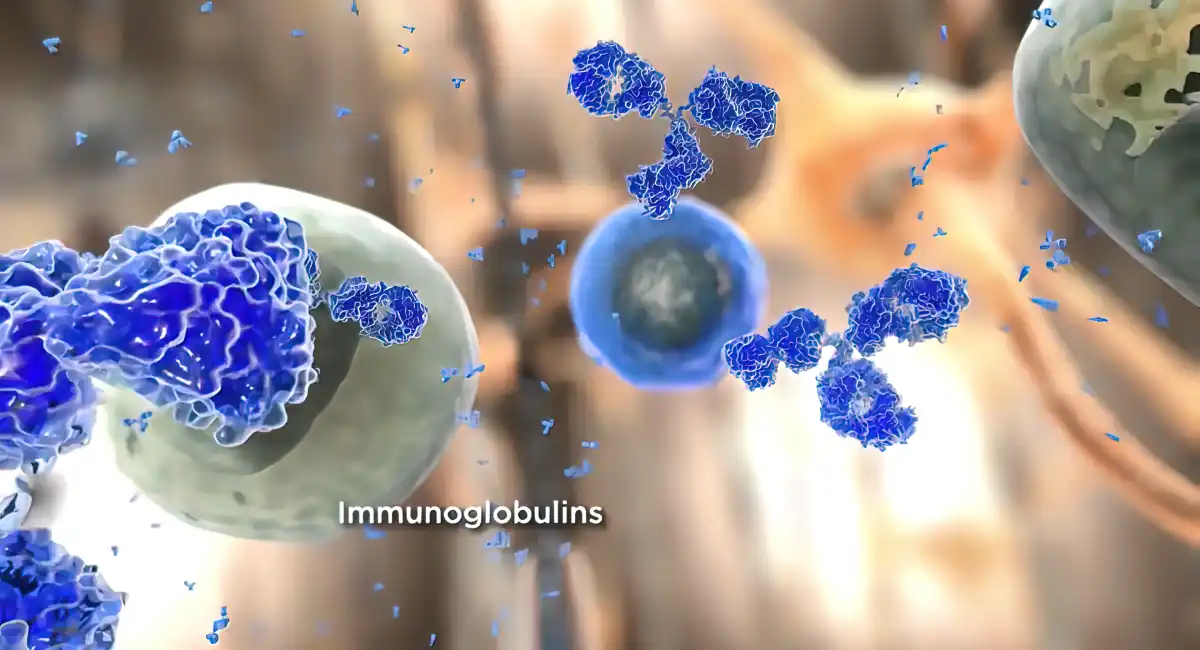
What is ELISA (Enzyme-Linked Immunosorbent Assay)?
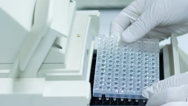
ELISA is a robust tool for detecting and quantifying proteins in complex study samples. This method can analyze proteins immobilized on a microplate well via specific antibodies. ELISA assays typically use 96 or 384-well plates that passively bind proteins and antibodies. This immobilization of target analytes and antibodies makes designing and performing ELISA assays easy and convenient. Once the reactants are immobilized on a microplate surface, separation of unbound sample components becomes easy. The ability to employ high-affinity antibodies and remove unbound sample components makes ELISA assays a critical tool to measure specific molecules in crude sample preparations.
Today, several ELISA assay variants are developed and employed in unique applications. However, all ELISA assay formats have the same following principles:
- Coating/capturing of analytes: direct or indirect antigen immobilization to the microplate (polystyrene) surfaces.
- Plate blocking: adding irrelevant molecules to occupy all unsaturated binding sites.
- Probing/detection: incubating the target molecules with specific antibodies and high affinity towards the target analytes.
- Signal measurement: detecting the generated signal through primary or secondary tags.
Horseradish peroxidase and alkaline phosphatase are the two commonly employed enzyme labels in ELISA methods. Besides, a library of substrates is available for conjugating with horseradish peroxidase and alkaline phosphatase. Selecting an ideal substrate depends on the available instrumentation and the required sensitivity.
Overview of ELISA Methods and Formats
 Capture antibody12Target
antigenEnzyme labelled
detection antibody3Substrate4
Several formats of ELISA methods are used in biomedical and life science applications. These ELISA assay methods fall into direct, indirect, and sandwich assays. The primary step is antigen immobilization by direct adsorption to microplates or indirectly through a capture antibody attached to the microplate. The antigen of interest is detected directly via a labeled primary antibody or indirectly using a secondary labeled antibody.
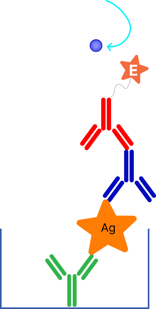
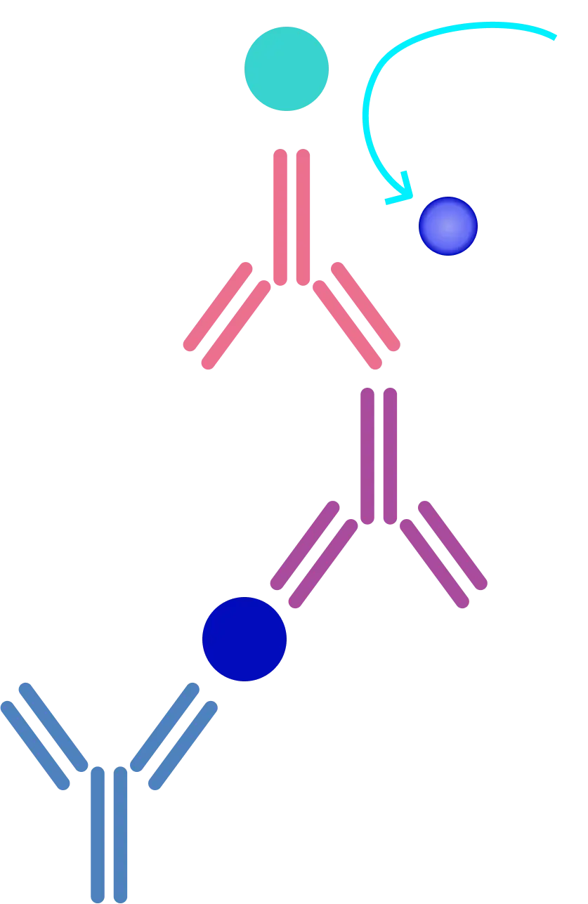
Today, ELISA labs predominantly use sandwich assay formats for immobilizing and detecting the target antigen. This assay format is termed sandwich assay because the target analyte is sandwiched between two primary antibodies, each identifying a unique epitope of the target antigen. The sandwich ELISA method is largely used due to its specificity and sensitivity.
Direct and Indirect ELISA Methods
 SubstratePrimary
antibody
conjugate
Direct Assay
 SubstrateSecondary
antibody
conjugatePrimary
antibody
Indirect Assay
 SubstrateCapture
antibody
Capture Assay “Sandwich”
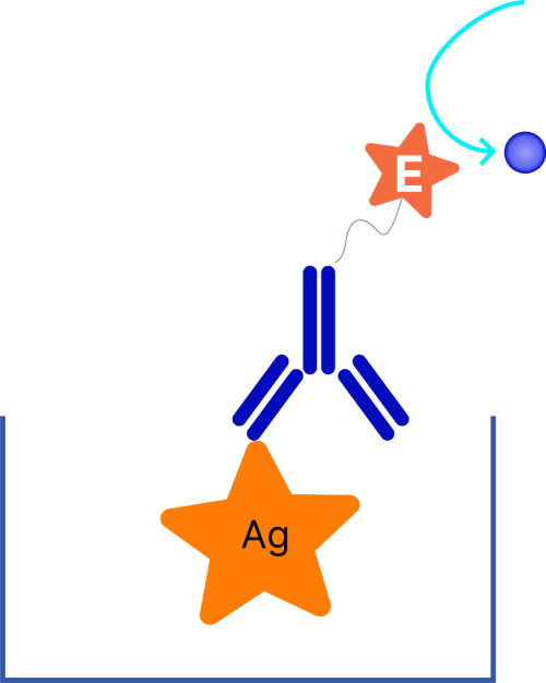
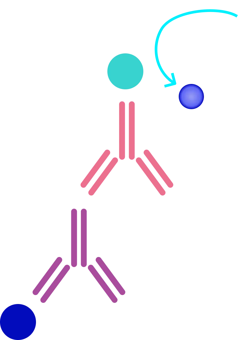
Direct and indirect ELISA are named based on the detection enzyme is conjugated with the primary antibody (Direct ELISA) or secondary antibody (Indirect ELISA). The direct ELISA method employs a primary antibody labeled with a reporter molecule that reacts with the target antigen. ELISA labs perform a direct detection method when the target antigen is immobilized on the assay plate or when using the capture assay method. Although the direct ELISA method is not widely used, it is found commonly in immunohistochemical tissue and cell staining applications. As direct ELISA assays use only one antibody, it has fewer steps and rapid protocol. Also, cross-reactivity is eliminated as secondary antibodies are not required.
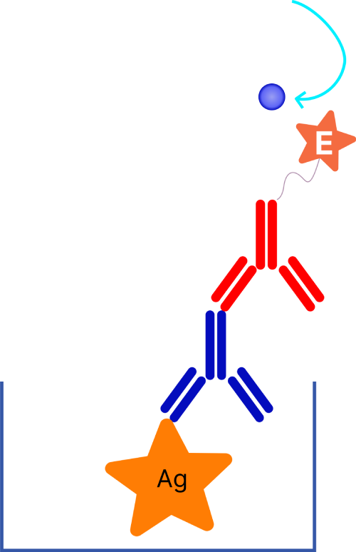
On the other hand, the indirect ELISA method employs a biotin-streptavidin complex or a labeled secondary antibody to detect the target antigen with the amplified signal. The secondary labeled antibody is specific to the primary antibody. One primary aspect of the indirect method is that as the primary antibody is not labeled, maximum immunoreactivity can be retained. Besides, indirect methods offer increased sensitivity compared to direct methods because the primary antibody contains multiple epitopes for the labeled secondary antibody, facilitating signal amplification.
Sandwich and Competitive ELISA Methods
In sandwich ELISA assay methods, the secondary antibody should only detect the primary antibody and not the capture antibody, or else the assay will lose its specificity. Ideally, this specificity requirement is met by using primary and capture antibodies from different host species, such as rabbit IgG and mouse IgG. Using cross-adsorbed secondary antibodies is ideal as it removes secondary antibodies that have an affinity towards the capture antibody. The primary advantage of the sandwich ELISA assay method is that it is specific and sensitive against the target antigen because two different antibodies are employed for capturing and detecting the target analyte. Besides, scientists can use different detection techniques for the same capture antibody.
 AgSubstratePrimary
antibody
conjugate
Direct Assay
 SubstrateSecondary
antibody
conjugate
Indirect ELISA
 SubstrateCapture antibody
Sandwich ELISA
 SubstrateInhibitor
Antigen
Competitive ELISA
Competitive ELISA
Competitive ELISA assay, also called blocking ELISA or inhibition ELISA assay method, determines the target antigen or antibody concentration in a sample by evaluating the signal interference in an expected signal output. The antigen or antibody of interest competes with the reference antigen or antibody. Competitive ELISA assays are commonly employed for smaller antigens with a single antibody binding site or epitope.
Competitive ELISA could adapt to the three formats of ELISA (direct, indirect, and sandwich) to form the three formats of competitive ELISA (direct competitive, indirect competitive, and sandwich competitive). The graph above shows the pattern of competitive ELISA when detecting antigens in the analyte.
Advantage and Disadvantage of Competitive ELISA:
| Advantage |
Disadvantage |
| Able to quantitate small molecules |
The same limitations (quality of antibodies, cross-reaction, non-specific binding, etc.) apply to competitive ELISA as the basic ELISA. |
| Lower sample requirement since it does not require extensive sample purification or concentration steps. |
| More robust since it is less sensitive to sample dilution and sample matrix effect (the presence of interfering substances in the matrix). |
Less sensitive than other ELISA types especially when analyte has high concentration. |
| More consistent and reproducible. There are fewer variations between interpolate duplicates and interpolate samples. |
More complicated since it needs to choose a proper reference antigen or antibody. |
| Maximum flexibility. It can adapt to direct, indirect, or sandwich ELISA. |
More challenging to interpret data because of the inverse relationship between signal output and concentration of analyte. |
Competitive ELISA is best used for (in the case of detecting antigen in the analyte):
- When only one antibody is available to detect the antigen of interest.
- Suitable for detecting small antigens and hormones that have only one antibody site and cannot be bound by two different antibodies, such as sandwich ELISA, due to steric hindrance.
ELISA Analysis – Understanding Detection Strategies
The final step in ELISA analysis is detection. Unless researchers use a fluorescent, electrochemiluminescence, or radioactive tag, the detection step involves an enzyme substrate. The enzyme reacts with the substrate to produce a detectable signal. Thus, the intensity of the generated signal after substrate addition is directly proportional to the total concentration of the target antigen bound to the assay plate. Enzyme conjugate antibodies are one of the most suitable and flexible detection methods for ELISA analysis as they can be used with different imaging tools such as chromogenic, chemiluminescent, and chemifluorescent detection.
Colorimetric detection
Colorimetric substrates develop a colored output that grows over time relative to the enzyme in each assay well. Once a desired color product is developed, the absorbance is measured directly, or a stop solution is introduced to halt the reaction and present a fixed endpoint. Although colorimetric substrates are not as sensitive compared to other detection methods, they offer direct visualization of the output. Besides, colorimetric detection can be easily performed using standard absorbance plate readers that are readily available in most laboratories.
Chemiluminescence detection
Chemiluminescence reactions involve the generation of energy in the form of light. A common approach in chemiluminescence detection is using luminol with horseradish peroxidase and a peroxide buffer. The oxidized luminol generates an excited product that produces light as it goes back to the ground state. Light is only emitted during the enzyme-substrate reaction. Hence, as the substrate is exhausted, the signal stops. Chemiluminescence reactions are relatively more sensitive than colorimetric methods. However, signal intensity in chemiluminescence detection may vary with other substrates. For ELISA analysis involving multiple plates, this signal intensity issue may cause a problem as the signal may decay before reading that particular plate. Hence, scientists should optimize the method with specific substrates to avoid misinterpretation due to signal fading.
Chemifluorescent detection
Fluorescent substrates are uncommon as their detection requires a fluorometer to produce the exact excitation beam to generate a signal. Chemifluorescence detection is an enzyme-based approach where the generated product is fluorescent. The generated signal is measured by a fluorometer using appropriate emission and excitation filters. Similar to chemiluminescence, researchers can measure chemifluorescence reactions directly over time or stop the reaction by using an appropriate chemical solution.
ELISA Lab Services and Applications
Due to high sensitivity and specificity and relative ease of use, ELISA assays have been implemented in several applications and services. Let us explore ELISA assay in different biomedical and drug development domains.
Pharmacokinetics (PK) Study Using ELISA
Assessing the efficacy and safety of drug products is a primary prerequisite for successful preclinical and clinical trials. During the drug development phase, the distribution, metabolism, and elimination profile need appropriate monitoring. Hence, pharmacokinetic evaluations are critical for drug discovery and development. Pharmacokinetic ELISA assays can determine drug concentrations in study subject samples at a given period. Especially in the early clinical assessment stage, pharmacokinetic studies offer auxiliary information about appropriate doses of biomarkers for subsequent stages of clinical trials. Therefore, regulatory agencies such as FDA and EMA have released multiple guidelines for addressing issues related to pharmacokinetic evaluations.
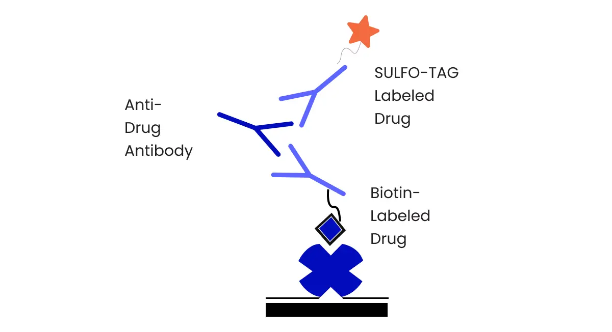
Often, researchers use sandwich ELISA assays for pharmacokinetic evaluations. In this setup, the target drug is captured and detected using two different antibodies. Moreover, a bridging ELISA assay, a unique sandwich assay format, is also used for pharmacokinetic studies. In this assay format, the antigen captured and detected by two different antibodies acts as a bridge for these two specific antibodies.
ADA Assay & Neutralizing Antibody Assay
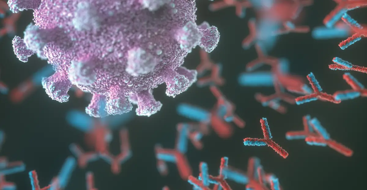
Monitoring and detecting anti-drug antibodies in samples is crucial for evaluating immunogenicity, addressing safety issues, and ensuring efficacy. This approach is critical for personalized medicine, where patients are identified to deliver alternative treatment or require dose adjustments.
Anti-drug antibody (ADA) assays detect ADAs binding to the target drug product. Several factors can generate anti-drug antibodies, including the host, drug product, and the study protocol and design. The impact of anti-drug antibodies may range from non-influencing effects to detrimental and life-threatening situations. Furthermore, assessing the neutralizing capacity through neutralizing antibody assays can help determine the magnitude and type of impact.
Researchers use several formats (direct, indirect, competitive, bridging, etc.) of the ELISA assay method for immunogenicity testing. The selection of assay formats depends on inherent drug properties and testing capacities of laboratories. The primary workflow of immunogenicity assessment has a multi-tiered approach, which begins with initial screening for positive samples, followed by confirmatory and titration tests. Additional testing, such as testing neutralization and isotyping, is conducted as needed.
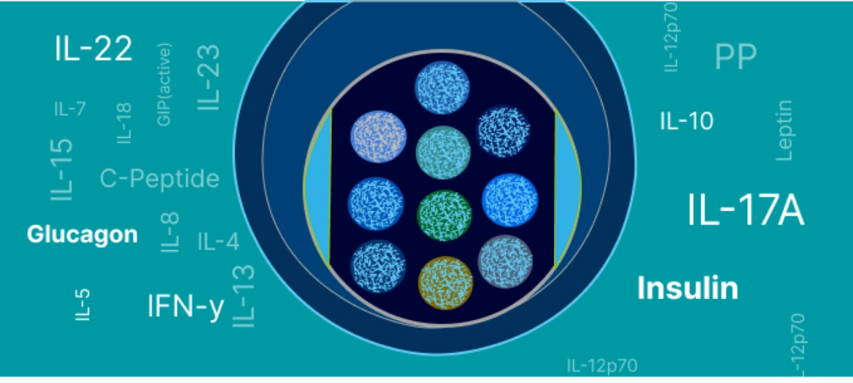
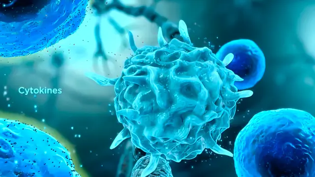
Cytokine Analysis & Biomarker Assay
Inflammation and cytokine analysis are complex endeavors. Cytokine and chemokine systems are critical in disease and medical conditions, including infectious diseases, autoimmune diseases, and cancer. Several ELISA methods, such as cytokine and biomarker assays, are available. Researchers employ these ELISA methods to measure levels of specific inflammatory markers and gain valuable information about the underlying mechanisms of disease and normal cellular functioning.
ELISA assays are user-friendly. They have reliable assay procedures, sample preparation, and data analysis protocols. ELISA methods are compatible with multiple sample types such as serum, blood, plasma, tissue lysates, etc, offering flexible testing alternatives. Additionally, sensitive ELISA assay formats are available to detect and quantify cytokines and other inflammatory markers present at low levels in biological samples. These assay formats offer greater precision and accuracy and allow a comprehensive assessment and interpretation of study data.
Cell-Based Assays Using ELISA
ELISA assays are often employed to quantify antigen or antibody binding activity. They are implemented to exploit the binding activity of antigen-antibody interactions and use it to generate and measure a spectrophotometric response. Cell-based ELISA assays are convenient, sensitive, high-throughput, and lysate-free. They can measure relative cellular protein phosphorylation. Besides, cell-based ELISA assays can monitor the impact of inhibitors, activators, or therapeutic interventions on phosphorylation.
Cell-based assays can evaluate several cellular biochemical processes. Hence, scientists now culture multiple cells and study their signaling, stimuli, and internal chemical processes. Cellular functions studied with cell-based ELISA assays include receptor binding and activation, ligand internalization, and cell signaling. Assessing these processes using cell-based ELISA assays offers a more detailed understanding of cellular functions in various environmental settings.
Bioanalytical Assay Validation
Validated ELISA methods for quantifying drugs, biological products, and biomarkers in complex biological matrices are crucial for successful preclinical and clinical studies. Bioanalytical assay validation instills confidence in the generated output and provides data to support the efficacy and safety of drug products.
Today, regulatory agencies such as the US FDA and EMA have clearly defined characteristics to validate bioanalytical methods, including ELISA assays developed for toxicological, diagnostics, basic, and applied research. Hence, scientists and drug developers should follow these guidelines as adhering to these recommendations is necessary for the results to be satisfactory and acceptable worldwide.
Advancements in ELISA Techniques and Technologies
Technological advancements in ELISA assay methods have led to more innovative and precise techniques, significantly improving the specificity and sensitivity of ELISA methods to detect and quantify proteins in complex study matrices. Let us explore some of these advancements in ELISA methods and technologies.
Integration of nanotechnology
Today, ELISA methods employ nanoparticles to load biomolecules onto the surface and artificial enzyme mimics that make these methods more stable and sensitive for detecting proteins. These mimics have improved the performance of techniques such as biosensors and proposed a new avenue in research and diagnostics. Besides, the application of quantum dots in ELISA analysis offers high stability and adjustable light emission, improving sensitivity and facilitating multiplexed assessments. Magnetic microfluidic ELISA systems can reduce the ELISA manual operation and save process time.
Multiplexed ELISA methods
Novel microarray ELISA assays detect multiple analytes in a single reaction volume, making them ideal for evaluating complex medical conditions such as neurodegenerative disorders. Due to high throughput abilities enabled by AI technologies and internet-of-things, scientists can now assess a spectrum of agents and proteins with greater accuracy and precision.
Automation in ELISA assays
Automated ELISA methods combine robotic handling along with multicolor sensing. It offers sensitivity, speed, and automation in diagnosing several diseases and disorders. Automated ELISA systems have enabled diagnostics and testing outside conventional laboratories. Moreover, incorporating microfluidic systems, automation, and nanotechnology could increase the sensitivity and specificity of current ELISA methods.
Optimizing ELISA Assays for Research and Clinical Diagnostics
Once an ELISA method is developed, assay optimization is needed to enhance its performance. The most critical aspect of ELISA optimization is assessing different dilutions of samples, antibodies, and buffers. Although each step of ELISA optimization is unique, two separate assay components can be optimized simultaneously using a checkerboard titration.
A thoroughly optimized ELISA method leads to adequate troubleshooting measures. For example, high background noise may be due to several factors such as insufficient washing, substrate exposure to light before use, incorrect standard curve dilutions, extended incubation period, etc. A highly optimized ELISA method ensures the availability of a reliable tool for subsequent analysis and validation protocols.