Scientific Significance of ELISA Validation
The need for assay validation in clinical diagnostics and pharmaceutical research
Validated ELISA assays for quantitative assessment of analytes such as biomarkers, drugs, and biological products in complex biological matrices, including blood, serum, plasma, or urine, are critical for successful clinical, non-clinical, and pharmacological studies. Validated ELISA assay provides crucial data to support the safety and efficacy of drugs and biological products. A thorough ELISA assay validation instills confidence in the generated data by addressing specific aspects of bioanalytical testing, such as:
- Understand whether the assay measures the target analyte. For example, identify whether something interferes with the assay measurement and is the ELISA assay selective or specific for the target analyte.
- Estimate assay variability to assess the precision and accuracy of the ELISA method.
- Identify the assay range that generates reliable data. For example, identify the upper and lower limits of quantitation to assess assay sensitivity.
- Impact of sample handling, collection, and storage on the accuracy and reliability of the generated data. For example, identify sample collection steps and storage parameters such as duration and temperature.
Regulatory compliance (FDA, EMA) and the role of Good Laboratory Practices (GLP) in ELISA validation
Today, abundant literature on immunochemical methods is available. Besides, regulatory agencies such as the US FDA and EMA have clearly defined parameters for validating bioanalytical methods. These guidelines also apply to ELISA validation developed for toxicology, diagnostics, and basic or applied applications. Hence, recommendations suggested for validating ELISA assays by these regulatory bodies are essential for generating satisfactory measurements that are accepted worldwide. ELISA validation based on these regulatory recommendations involves characterizing the following parameters:
- Sensitivity
- Specificity
- Linearity
- Precision
- Accuracy
- Robustness
Most importantly, the acceptance criteria for each of these parameters should be based on the intended application of the ELISA assay.
Consequences of poorly validated assays
A poorly validated ELISA assay can lead to several potential errors. Hence, incorporating best practices and following regulatory guidelines becomes critical during ELISA assay validation. A significant consequence of poor ELISA assay validation is the generation of false positive and false negative results.
Common issues during ELISA validation, such as insufficient plate washing or too much detection reagent, may yield a high signal, which, in turn, can generate false positive results. Besides, cross-reactivity or contamination may increase the background signal in an ELISA assay. Such a high amount of background generates false positive or false negative results and impacts the accuracy of the generated data. Additionally, maintaining lot-to-lot consistency is critical because failure to do so may lead to false negatives and false positives.
Core Validation Parameters in ELISA Assay Validation
Let us turn our attention to the core parameters in ELISA assay validation.
Precision and Reproducibility
|
Intra-Assay Precision |
Inter-Assay Precision |
| Sample |
1 |
2 |
3 |
1 |
2 |
3 |
| n |
20 |
20 |
20 |
20 |
20 |
20 |
| Mean(pg/ml) |
175 |
383 |
582 |
178 |
379 |
599 |
| Standard deviation |
7.9 |
16.6 |
20.9 |
10.2 |
21.5 |
28.7 |
| CV(%) |
4.5 |
4.3 |
3.6 |
5.8 |
5.7 |
4.8 |
Intra-assay precision
Intra-assay validation demonstrates the reproducibility among individual wells on an assay plate. Intra-assay validation ensures that samples in each well will give comparable results.
Inter-assay precision
Inter-assay precision confirms reproducibility among ELISA assays performed on different days. Typically, inter-assay precision is less than 10%. This range ensures the consistency of generated results over an extended period and among different assay kits.
Sensitivity and Lower Limit of Detection (LLOD)
Sensitivity is the identification and detection of analytes at the lowest level from the background. This detection is also called the lower limit of detection. However, it is critical to remember that the lower limit of detection differs from the lower limit of quantification nominal. Researchers determine the limit of detection using the standard deviation of the sample blank and the linear curve slope.
The goal in ELISA assay development is a high signal-to-noise ratio while generating optimal responses. The sample matrix may have interfering elements that may influence the assay response. Hence, researchers often design spike and recovery experiments to achieve a high signal-to-noise ratio and adequate assay sensitivity.
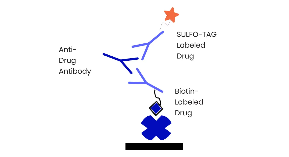
Specificity and Cross-Reactivity
Specificity refers to the ability of an ELISA assay to differentiate the target molecule from other matrix components such as endogenous compounds, physicochemically related compounds, isoforms, and variants of the target analyte. In other words, specificity confirms that the target analyte is detected without cross-reactivity with related molecules. Cross-reactivity can cause inaccurate quantitation and generate false positive results. Hence, researchers test a panel of associated substances to evaluate cross-reactivity and ensure that the target analyte is detected.
Studies employing ELISA assays confirm specificity by using increasing concentrations of related endogenous components in the sample and measuring the total amount of target macromolecules within the working assay range. Additionally, specificity can be tested using competitive ELISA assays or assessing negative immunization control samples.
Linearity and Range
Linearity is the capacity of an ELISA assay to generate data by estimating a direct proportion within the assay range. Assay linearity is expressed relative to the calculated analyte concentration. Linearity is a graphically represented function of values and can be described as an assay’s ability to demonstrate data that is directly proportional to the analyte concentration in the study sample. However, all analytical responses may necessarily not always be linear. Hence, non-linear data needs to be mathematically modified by using algorithms.
Scientists determine assay range from linearity. The generated data should fall within the assay range and satisfy the criteria for accuracy and precision. Moreover, an assay range is capped by the upper and lower limits of detection.
Accuracy and Recovery
The accuracy of an ELISA relates to the closeness of individual values obtained through nominal concentration. Biological samples are precious. Hence, in practice, the primary step is preparing quality control samples that are standardized with the reference material. These standardized quality control samples are used to determine different validation parameters. Generally, scientists evaluate accuracy using samples that are spiked with the analyte of interest.
Biological samples such as plasma and serum have interfering elements that could impact the quality of an ELISA assay. Hence, recovery experiments are needed to evaluate the differences in matrices that may impact analyte detection.
Robustness and Stability
Evaluating assay robustness involves studying the impact of minute unintentional errors on the qualitative and quantitative characteristics of analysis. These errors correlate to assay parameters such as buffer temperature, sample incubation period, number of wash steps, incubation temperature, color development time, secondary antibody incubation period, etc. Any variations in environmental factors or assay reagents impact the robustness of a bioanalytical method. Hence, robustness and stability testing are critical to ensure assay performance over time and demonstrate the reliability of an ELISA assay despite minor changes in performance.
Practical Steps for Validating an ELISA Assay
Optimization of Reagents and Protocols
Once an ELISA assay is developed, scientists perform optimization protocols to improve assay performance. Below are some practical optimization steps for individual components of an ELISA.
The concentration of capture antibodies
- Make different capture antibody concentrations in the coating buffer
- Evaluate strong signal versus low background
- Use the same volume of individual concentration in the ELISA plate
- Choose a high-affinity antibody and titrate the concentration of antibodies to optimize concentration for the best signal-to-noise ratio
Blocking buffer
- Make different blocking solutions.
- Test diverse blocking buffers and different concentrations of blocking buffers to cover any spots on the plate well and prevent non-specific binding.
- Optimize blocking buffer.
Sample concentration
- Match the standard diluent to the sample matrix. If the exact sample matrix is not possible, test different solutions of standard diluent.
- Have a good sample dilution linearity and standard curve dynamic range.
- In case of a poor dynamic range for the standard curve, select a different diluent.
Signal detection
- Substrate selection should depend on the total antigen in the study sample and its detection with appropriate instruments.
- A substrate is appropriate when the target antigen is detected across a dynamic range. For antigens below the detection threshold, select a sensitive substrate.
Thorough washing
- To remove any unbound reagent, ensure thorough washing throughout the whole process.
Calibration Standards and Parallelism
Calibration of the assay using known reference standards
A standard curve confirms the quality of an ELISA assay and makes the operation protocol acceptable for subsequent testing. In ELISA quantification, determining the quantity of unknown samples requires a set of reference standards with known quantities. These reference standards require testing at different concentrations ranging from undetectable signals to maximum output. The yielded data for reference standards allows for matching a model and generating a standard. Generating a standard curve makes it relatively easy to identify where the unknown sample lies on the curve and interpolate that specific value.
A standard curve is developed using serial dilutions of the reference standard with known concentration spanning the entire standard curve range. The range of a standard curve is set to be close to the concentration range of the target analyte in the sample.
Importance of parallelism in ensuring comparability across dilution series
Parallelism confirms whether samples with high concentrations of endogenous analytes offer the same level of detection after dilutions in the standard curve. This confirmation signifies the differences between the standard calibration analyte and the affinity of antibody binding to the endogenous analyte. Generally, parallelism is performed while validating test samples.
Parallelism uses samples with high spiked standard analyte concentration and high endogenous analyte levels. However, using highly concentrated samples is essential in parallelism studies as the true parallelism value cannot be detected by using concentrations that are close to the lower end of detection.
The absence of parallelism indicates the presence of matrix effects or differences between the nature of protein used in reference standard to native protein. Either case requires assay optimization, which includes the choice of different reference proteins, different antibodies for assay, optimization of incubation time, or using another assay buffer.
Validation Experiment Design
The primary goal of ELISA assay validation is to prove that the developed assay is suitable for analyzing the study sample. Regulatory agencies have put forth recommendations and guidance for designing validation experiments and developing reproducible protocols. Following are some critical aspects highlighted by the US FDA for bioanalytical assay validation.
- Perform full assay validation for new methods developed for analyzing a new drug compound, metabolites, or biomarkers.
- Perform a full assay validation for any modification to an existing ELISA assay where a new analyte or metabolite is added.
- Establish a detailed description for the bioanalytical assay before beginning method validation. This description should include protocols that control critical assay parameters from the time of sample collection to the time of its assessment to minimize errors in assay measurement.
- Document and report all tests performed to draw conclusions or make claims about assay validity.
- Validate the test for each analyte in the study biological matrix.
Additionally, sponsors should characterize and document critical reagents such as reference standards, labeled analytes, matrices, and antibodies. Assay validation is crucial when critical reagents are changed, such as switching to a new reagent or lot-to-lot changes. For example, any change in the antibodies for detection reagents will require evaluation of the binding process, verification of the assay performance using quality control and standard curve, and evaluation of cross reactivity. Besides, the use of multiple kit lots in the same study will require consideration of lot-to-lot comparability and variability for critical reagents.
Quality Control Measures in ELISA Validation
Importance of Controls
Verifying quantitative results is critical before reporting. This verification requires an established quality control system for all qualitative and quantitative analyses. When establishing such a quality control system, laboratories should train staff, assign responsibilities, set policies, and ensure the availability of all necessary resources.
Positive and negative control samples are necessary for several qualitative and quantitative assessments, including protocols that use specialized reagents and experiments with endpoints such as color change or agglutination. Generally, each test run should include these controls. The use of positive and negative controls has multiple applications, such as validation of a new lot of reagents, assessing storage and testing area temperatures, and evaluating the system when a new team member runs an experiment. Following are some considerations laboratories and sponsors should keep in mind when using traditional control samples in qualitative and quantitative assessments.
- Use positive and negative controls.
- Keep the testing protocol similar to the study sample and control materials.
- Select positive control samples with cut-off values close to the test.
- For agglutination protocols, include a weak positive control, a negative control, and a stronger positive control.
Lot-to-Lot Variability
Different assay lots require testing to ensure consistent sensitivity, low background detection, and linear standard curves. ELISA assay kits developed on a lot-to-lot basis require confirmation that the data produced at different intervals by independent users are comparable. This comparison is attained by studying lot-to-lot differences and verifying whether results obtained from the new kit correlate with results from old ELISA kit batches.
Lot-to-lot comparison of new and old ELISA assay kits is performed before the old lot expires. Lot-to-lot variability is assessed by evaluating positive samples using the old and new ELISA kits on the same day. Same-day assessment of lot-to-lot variability is highly accurate as performing tests on the same day avoids the differences that may originate from the distinct stability of the target analyte in the study matrix. A manufacturer compares the standards of both lots to a master calibrated reference standard developed against a purified material that is used as an internal control. Consistent control values of the master reference standard ensure reproducibility in different kit lots.
Common Challenges and Troubleshooting in ELISA Validation
While developing and validating ELISA assays, there are several instances where potential problems may occur and impact assay results. Let us dive deep and understand the most common issues.
Handling Matrix Effects
ELISA quantification using biological samples faces problems due to matrix effects. The matrix effect can be due to several sample components, such as the interaction between endogenous elements, including carbohydrates, metabolites, and phospholipids, or the interaction between the target analyte and the matrix component, such as plasma proteins and other components for example buffer salt, pH or additives. Matrix effects lead to errors (either falsely depressed or falsely elevated analyte levels) in sample readings.
There are several ways to tackle matrix effects. One of the easiest and preferred approaches is to overcome sample matrix interference by diluting the sample into a more assay-compatible buffer. The ideal diluent is the same material used to prepare the standard curve. Dilution is only used when the sample has an analyte that remains within the analytic range (not fall below LOQ) after dilution. In this case, Minimum Required Dilution (MRD) can be established.
When dilution is not an option either because it does not help with resolving the problem or it does not fit for sample detection range and when the problem is most likely the cause is buffer salt, since it is small molecular weight, could consider using commercially available desalting columns and adding additional volume of buffer during purification to resolve this issue.
Another approach is neutralizing samples to overcome pH issues. In general, a pH between 7.0-7.5 is ideal for ELISA assay. Adding buffering concentrated could correct the sample pH.
Modification protocol by decreasing sample size, longer incubation time, etc., could also help to improve the assay result caused by matrix effects.
Improving Sensitivity and Specificity
Let us consider possible causes and potential solutions to improve assay sensitivity and specificity.
Inappropriate storage: different reagents may have unique storage requirements. Hence, follow the recommended conditions while storing reagents.
Inactive detection reagent: confirm the expected activity of the reporter enzyme.
Insufficient target: reduce the sample dilution or concentrate the sample.
Incorrect plate reader settings: always check whether the plate reader is set at the right excitation/emission wavelength or absorbance wavelength in fluorescent detection.
Assay not sensitive enough: change to a sensitive detection system or assay type. Moreover, one may raise the temperature or lengthen the incubation time to increase assay sensitivity.
Interpretation of Results
While interpreting ELISA results, specific initiatives are essential, and multiple parameters should be considered before and after an ELISA assay. Let us understand the necessary factors required before and after running an ELISA assay for robust interpretation of results.
Before running an assay
Use replicates
Each sample and standard should be run in replicate to assess the extent of error. After running samples in replicates, researchers can calculate the standard deviation, average, and coefficient of variation to instill confidence in the pipetting technique.
Have a standard curve on every assay plate.
Each ELISA assay varies depending on the pipetting, incubation, operator, and temperature. Running a standard curve on each assay plate is a best practice to accommodate different variables affecting the ELISA assay.
Use positive control
Having a control sample with known concentrations on each plate will help confirm whether an ELISA assay was executed successfully.
Run blank samples
Blank samples can ensure accurate optical density readings as they allow the subtraction of background noise from the remaining data points.
Dilute samples
Sample dilution can be considered, especially when facing matrix effect problems since it could help samples stay within the linear range and get accurate results.
After running an assay
Use a four-parameter algorithm.
Ideally, develop a standard curve using curve fitting software such as a four-parameter algorithm.
Subtract background absorbance
Do not forget to use blank samples and subtract the background absorbance from the readings. If the blank sample reading is high and the signal-to-noise ratio is relatively low, it indicates there are issues in the assay.
Account for dilution factors
Once the absorbance of an unknown sample is read, multiply the calculated concentration based on the linear standard curve with a dilution factor.
Calculate the standard deviation average and coefficient of variation
After considering the dilution factor and blanks, one can calculate the concentration of the unknown sample with the standard curve. Then, in the case where replicates were used, assess the results for the standard deviation, average, and coefficient of variation for the final assay results.
Real-Life Applications of ELISA Validation
ELISA assay validation translates into several applications across different domains, including oncology, the food industry, and immunodiagnostic.
Oncology
Highly sensitive diagnostics for cancer patients offered during the early stage are a crucial indicator for patient survival. However, identifying oncology biomarkers is one of the most challenging components of target analytes. Today, advancement in ELISA validation and development has resulted in the availability of several ELISA-based techniques for testing early stages of cancer, including breast and ovarian cancer.
Food industry
ELISA has a significant role in the advancement of the food industry. Today, ELISA assays are the primary platform for detecting food allergens, such as components present in eggs, almonds, walnuts, peanuts, and milk. The application of food allergen testing primarily benefits from the high sensitivity of ELISA, which helps detect potential food allergens present at low concentrations. Most importantly, ELISA-based assays can evaluate specific substances, such as egg whites and oils, which other methods, such as PCR, cannot detect because they do not contain DNA.
Immunodiagnostic application
In the field of immunodiagnostics, ELISA has proven its ability to detect several disease types in animals and humans. Also, ELISA assays are increasingly being employed in plant pathology. Today, multiple commercial ELISA assay kits are available for diseases such as HIV, Ebola, influenza, dengue fever, West Nile virus, and Lyme disease, even the most recent COVID test kit.
Advanced ELISA Validation Techniques
Multiplex ELISA Validation
Multiplex immunoassays have several advantages over singleplex immunoassays. However, successful multiplex assays depend on thorough and careful development and validation. Often, ELISA assay development and validation are limited by matrix interference, cross talk, lot-to-lot variability, and availability of matched antibody pairs.
The quality and performance of a multiplex ELISA assay depends on the matched antibody pairs. Antibodies validated on a singleplex platform do not necessarily have the same qualities in a multiplex system. Hence, scientists should validate antibody pairs on the same platform used in the final assay to ensure adequate method performance.
The variability observed among different reagent lots can impact ELISA assay validation. Researchers should document and track variability observed in the raw materials, such as antibody pairs and buffer formulation, and optimize variations to reduce the impact on the finalized assay model. Besides, robust ELISA assay validation strategies, such as ordering raw materials from the same vendor and lot whenever possible, should be in place to control lot-to-lot variability.
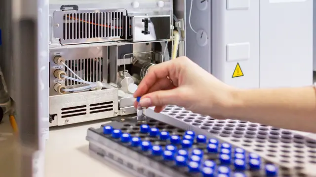
Automation in ELISA Validation
ELISA assay validation can be time-consuming and complex. Besides, it is open to human error, making it an ideal method for automation.
Automation in ELISA validation will improve assay accuracy and reduce random and systematic errors. Besides, automated systems reduce variability caused during work flows when separate individuals perform an experiment or an assay is conducted in different laboratories.
Automation ensures that the same protocol is followed every time, standardizing the entire ELISA validation protocol, eventually increasing reproducibility and reducing variability. Most importantly, automated systems can perform tasks more quickly and accurately. This quickness increases the speed and efficiency of ELISA validation, making more time for researchers to focus on other crucial tasks.