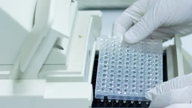
Basics of ELISA – Principles and Mechanisms
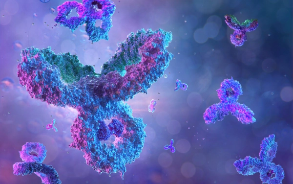
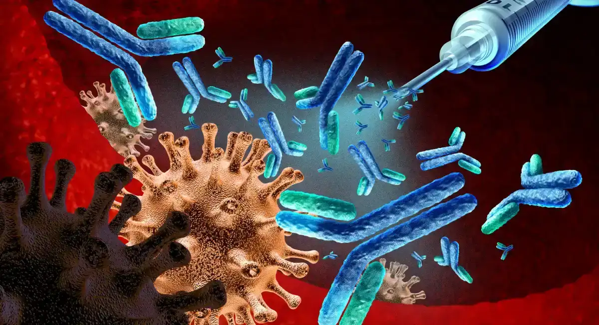
Binding interactions between antigen and antibody
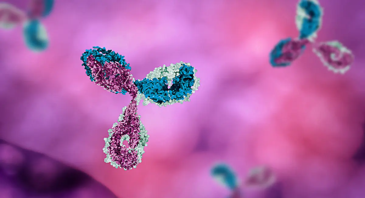
Antigen and antibody are substantial elements that determine the specificity and sensitivity of an ELISA assay. The antigen with good stability and purity can enhance the capturing capacities of antibodies, eventually increasing assay sensitivity.
The 3D structure of an antigen binding site in the Fab section of an antibody determines the specificity and strength of an antigen-antibody interaction. The stronger the antigen-antibody interaction, the lower the antigen concentration can be detected. A critical competing element in this interaction is the cross-reactivity of an antibody to other components in the serum rather than the target antigen. Depending on whether monoclonal or polyclonal antibodies are used, polyclonal antibodies comparatively possess higher cross-reactivity than monoclonal antibodies.
In sandwich ELISA assays, researchers can use polyclonal or monoclonal antibodies as capture and detection antibodies. Monoclonal antibodies are inherently mono-specific towards a single epitope and allow detection and quantification of minute alterations in the target antigen. On the other hand, a polyclonal capture antibody can gather as much antigen as possible. Once a polyclonal capture antibody entraps a majority of antigens, a monoclonal antibody can then be used to improve assay specificity.
A crucial aspect of sandwich ELISA development is choosing a capture and detection antibody system that recognizes two distinct non-overlapping epitopes. Once an antigen attaches to the capture antibody, that detection antibody epitope should not be altered or obscured. A matched antibody pair is a set of antibodies that detect different epitopes on the same protein antigen. With matched antibody pair, capture and detection antibody systems can bind simultaneously and do not influence epitopes of each other. Besides, matched antibody pairs could be used to capture and detect a single antigen in a sandwich ELISA or related immunoassay.
Types of ELISAs (Direct, Indirect, Sandwich, and Competitive)
 SubstratePrimary
antibody
conjugate
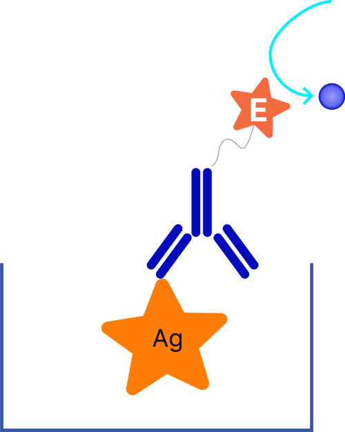
Direct Assay
 SubstrateSecondary
antibody
conjugatePrimary
antibody
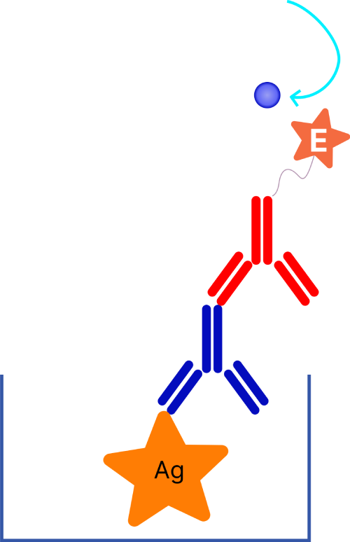
Indirect Assay
 SubstrateCapture
antibody
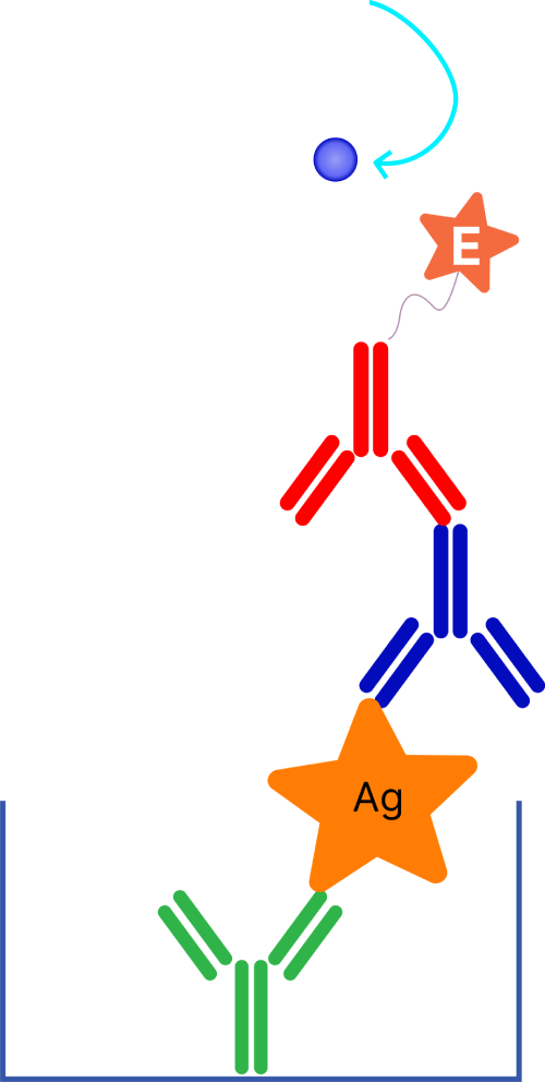
Capture Assay “Sandwich”
The four primary ELISA assay formats include direct, indirect, sandwich, and competitive assay. The sandwich, direct, and indirect formats differ based on the protein initially immobilized to the solid surface and the detection system. On the other hand, competitive ELISA assays are slightly different in their working principle, and they are readily adapted to all other three assay formats. However, in all formats, washing steps are critical to remove unbound protein, and blocking of wells is necessary to minimize non-specific binding. Let us understand all these ELISA assay formats in brief.
Direct ELISA assay
Direct ELISA assay was the first format developed and is one of the easiest ELISA assays. A target antigen sample is immobilized to the assay well surface where the protein binds with high affinity. An antibody conjugated to an enzyme or other detection molecule detects the target antigen.
Indirect ELISA
The primary difference between direct and indirect ELISA is the detection process, which has two steps. It involves an unlabeled primary antibody binding to a target antigen and a labeled secondary antibody against the host species of the primary antibody binding to the primary antibody for detection. This two-step process offers greater sensitivity as multiple secondary antibodies can bind to the primary antibody and produce the amplified signal for detection.
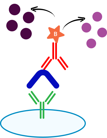
Sandwich ELISA
Sandwich ELISA has this specific name because the target antigen is sandwiched between two antibodies. A primary capture antibody immobilized on the plate surface binds to the target antigen, while a detection antibody binds to the same antigen at a different epitope. Such a unique system of matching antigen-antibody pairs makes sandwich ELISA assays much more specific than direct or indirect ELISA formats. However, without a commercial or standardized sandwich ELISA, identifying a matched antibody pair requires adequate optimization.
Competitive ELISA
Competitive ELISA is also known as inhibition ELISA since the sample competes with a reference. Each of the previous three formats can be adapted to this particular type of ELISA. There are two ways for performing competitive ELISA: one way is immobilizing reference antigen on a plate, then sample antigen competes with reference antigen for binding a labeled antibody, and the signal detected is the level of reference antigen; another way is coating the plate with primary antibody, and the sample antigen will compete with labeled antigen. In either case, more antigens in the sample will get a weaker signal because of the competitive binding.
Importance of enzyme-substrate reactions in signal detection
The enzyme-substrate reaction is crucial for signal detection in ELISA assays. The antibody-enzyme conjugate is incubated with the corresponding substrate to generate a measurable product. This product gives data on the target antigen. A robust enzyme-substrate system ensures reliable detection of target antigens in an ELISA assay.
Enzyme-labeled antibodies are critical for generating ELISA results. Conjugating enzymes with antibodies involves a stable covalent bond between an enzyme and a monoclonal or polyclonal antibody specific to the target antigen. Several reporter enzymes, such as alkaline phosphatase and horseradish peroxidase, can be used in ELISA assays. Horseradish peroxidase is a glycoprotein enzyme that can be visualized through chromogenic reactions such as chemiluminescence. On the other hand, alkaline phosphatase is a hydrolase enzyme where the signal is measured using colorimetric substrate pNPP.
The intensity of the generated signal after substrate addition is directly proportional to the total antigen concentration present in the sample. The substrate for horseradish peroxidase is hydrogen peroxide. In this substrate reaction, TMB is the most common dye used to generate the desired signal. The substrate and dye for alkaline phosphatase is pNPP. Generally, horseradish peroxidase has a faster reaction time than alkaline phosphatase and is the preferred enzyme-substrate system.
How Do You Develop an ELISA Assay Step by Step?
-
1. Coating
-
2. Analyte capture
-
3. Detection antibody
Although several ELISA assay variants are developed and employed in unique applications, they all have the same four primary elements. Let us explore these critical steps in ELISA assay development.
- Coating/capture: direct or indirect capture of target antigens onto the surface of microplate wells.
- Plate blocking: addition of other proteins or molecules to cover unsaturated binding sites of the assay well surface.
- Probing/detection: incubating with direct or indirect (2nd Ab) antibodies specific to the target antigen.
- Signal measurement: detecting the generated signal through direct or secondary labeled antibodies.
The fundamental step of establishing an effective ELISA assay is immobilizing the target antigen. This step is achieved indirectly through a capture antibody or directly via adsorption. The second step includes detecting the target antigen directly through labeled primary antibodies or indirectly via labeled secondary antibodies.
The sandwich ELISA assay format is one of the most widely used formats, which employs indirect immobilization and detection to identify the target antigen. This format is called sandwich assay because the target analyte is sandwiched between two antibodies, each detecting a unique epitope. The sandwich ELISA assay format is employed highly due to its specificity and sensitivity.
The following sections now dive deep into the particular nuances of different steps of ELISA assay development.
What Is the Best Coating Strategy for ELISA Method Development?
Choosing the correct coating strategy is essential for ELISA method development, ensuring optimal binding of antibodies or antigens. Although researchers use 96/ 384 well microplates made of polystyrene/polyvinyl/polypropylene, multiple solid phase materials are available during ELISA method development.
The approach used to immobilize proteins determines the core properties of an ELISA assay. In some scenarios, immobilization of proteins may lead to a complete or partial loss of their activity due to structural deformation or random orientation. Hence, to retain its entire biological activity, researchers should attach the proteins to the plate surfaces without altering their function and conformation. Generally, an ideal immobilization strategy depends on the chemical and physicochemical properties of the plate surface and the target protein. Different immobilization techniques are available. However, they primarily follow three mechanisms: covalent, physical, and bioaffinity immobilization.
How Do You Select the Right Antigen and Antibody for ELISA Assay Development?
Selecting high-affinity antigen-antibody pairs is crucial for successful ELISA assay development. Antigen and antibody pairs are critical determining factors for assay specificity and sensitivity. The stability and purity of an antigen affect the assay performance. Besides, high antigen purity enhances the capturing capacity of an antibody, eventually increasing assay sensitivity.
Researchers can use polyclonal or monoclonal antibodies to capture and detect antigens in sandwich ELISA assays. Monoclonal antibodies are inherently specific toward a single epitope and can detect and quantify minute differences in the target antigen. On the other hand, a polyclonal antibody can capture as much of the target antigen as possible. Scientists can then employ a monoclonal antibody to detect the antigen and offer enhanced specificity to the sandwich ELISA assay system.
A crucial element in sandwich ELISA assay development is that the two antibodies must identify two different non-overlapping epitopes. Most importantly, once an antigen attaches to the capture antibody, it should not alter or obscure the epitope of the detection antibody. The detection and capture antibodies that do not impact each other and can bind simultaneously to the target antigen are called matched antibody pairs. These matched antibodies are ideal for a sandwich ELISA assay.
What Conjugate Strategy Works Best in ELISA Method Development?
The ELISA signal output depends on the enzyme-labeled antibody. Conjugating an enzyme with an antibody during ELISA method development involves forming a stable covalent bond between an enzyme and a monoclonal or polyclonal antibody. However, the primary aspect of this enzyme antibody linkage is that neither the active site of an enzyme nor the antigen attachment site of an antibody is functionally altered.
Several reporter enzymes, such as alkaline phosphatase and horseradish peroxidase, can be used to generate maximum retention of both protein and enzymes. Horseradish peroxidase can be visualized through chromogenic reactions. On the other hand, alkaline phosphatase is a hydrolase enzyme, and its activity is determined often through colorimetric substrates. However, horseradish peroxidase is relatively more stable than alkaline phosphatase.
How Can You Optimize Enzymes and Chromogens in ELISA Development?
Detection is the final step in an ELISA assay system. The intensity of the generated signal post substrate addition is directly proportional to the antigen captured in the assay well and bound to the detection reagent. Hence, enzyme and chromogen selection can significantly impact the sensitivity and specificity of ELISA development.
The most common enzyme-substrate combination is horseradish peroxidase with hydrogen peroxidase and alkaline phosphatase with para nitrophenyl phosphate. Moreover, the horseradish peroxidase and hydrogen peroxide combination employs five different dyes, including OPD, DAB, TMB, 5AS, and ABTS. Besides, each of these enzyme-substrate and dye combinations has unique buffer systems and stopping solutions.
How Do You Troubleshoot ELISA Method and Assay Development?
ELISA method development includes several unique steps where potential issues may occur and influence the assay result. Hence, effective troubleshooting is essential for ELISA method development and ensuring reproducible and accurate results. The most common issues observed during ELISA assay development are related to:
- Poor standard curve
- Matrix effect
- Weak or no signal
- Poor reproducibility
- No color development or slow color development
- Low sensitivity
- Large coefficient of variation
- High background
- Positive data seen in negative controls
Let us consider some possible causes and troubleshooting tips for each of these issues observed during ELISA method development.
Poor standard curve
Possible cause: improper standard solution.
Corresponding solution: validate that dilutions are prepared correctly.
Matrix effect
Possible cause: matrix effects can be caused due to multiple components such as endogenous phospholipids, metabolites, carbohydrates, etc.
Corresponding solution: diluting the study samples can reduce the matrix effect. Use the same diluent that was employed for the standard curve.
Weak or no signal
Possible cause: too brief incubation period.
Corresponding solution: incubate the study sample overnight or follow the guidelines provided by the manufacturer.
Poor reproducibility
Possible cause: samples and kit controls were at different temperatures.
Corresponding solution: allow the study samples, kit controls, and sample diluent to come at room temperature. Generally, a larger volume of study samples will require an extended equilibration period. If a water bath is used to achieve equilibrium, ensure it is at room temperature.
No color development or slow color development
Possible cause: detection reagent is contaminated, too old, or utilized at the wrong pH.
Corresponding solution: use freshly prepared detection reagents. Reagents with contaminants, such as peroxidise and sodium azide, can impact the substrate reaction. Hence, avoid reagents that contain these preservatives.
Low sensitivity
Possible cause: incorrect plate reader settings
Corresponding solution: set the plate reader at the right excitation/emission wavelength or absorbance wavelength in fluorescent detection.
Large coefficient of variation
Possible cause: cross-well contamination.
Corresponding solution: ensure that the reagent does not touch the sealer when reusing these plate sealers. Besides, care is necessary when using the same tips for adding reagents.
High background
Possible cause: wells are contaminated or insufficiently washed.
Corresponding solution: increase the total number of washes or the soaking duration between washes. A sealer should also be used to avoid cross-well contamination. Moreover, multi-channel pipettes can help deliver the reagent without touching the plate.
Positive data seen in negative controls
Possible cause: use of too much antibody, causing non-specific binding.
Corresponding solution: always verify the recommended antibody concentration. If necessary, use less antibody.
Conclusion
ELISA assays are a robust technique for studying analytes in complex materials. However, ELISA method development requires an in-depth understanding of the target analyte, study matrix, and assay format. NorthEast Biolabs offers reliable ELISA services, specializing in ELISA method development. As industry leaders, we support every aspect of drug discovery and development. NorthEast Biolabs can develop robust ELISA assays and deliver accurate, reliable results in a cost-effective and timely manner.