Expertise
Services
Solutions
About Us
 About Us
 Overview
 Leadership
 Facility & Equipment
 Quality & Compliance
 Therapeutic Areas
 Why Us?
 Values
 Process
 Partners
 Careers
Sr. Scientist – Cell Based Assay
Associate Scientist, Central Laboratory Services
Business Development Associate
Sr. Scientist, LC-MS/MS Bioanalysis – Hamden, CT, USA
Sr. Scientist, ELISA Immunoassay Bioanalysis – Hamden, CT, USA
GLP Quality Assurance Manager
Sr. Scientist - Pharmacokinetics (PK)
Content Manager, Scientific and Regulatory Writing
Resources
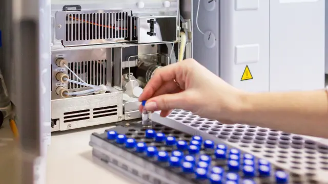
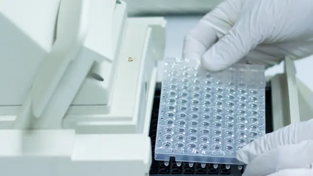
ELISA Testing Service, ELISA Assay Validation & Development by PhD Scientists at 20+ Year, FDA-Inspected Lab
De-Risk Your ELISA Validation & Testing, Non-GLP to GLP.
Brief PhDs on your analyte & samples.- ELISA Testing For MAb, ADC, Bispecific, Cytokine, Protein, Peptide, Oligo, Small Molecule, CGT
- Non-GLP, GLP ELISA Validation For PK/PD, ADA/NAb, Activity/Potency, Biomarkers, Viral Titers, Vaccines
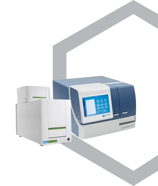
- Preclinical, Clinical ELISA Services Via Absorbance, MSD, Luminescence, Fluorescence, TR-FRET, AlphaLISA
- ELISA Analysis Using Proprietary, Commercial Antibodies, Kits In Serum, Plasma, Urine, Saliva, CSF, Tissue, Cells
Regulated And Inspected By:

The Scientific Team Behind Your Bioanalytical Service
20+
Years In Bioanalysis
700+
Sponsor Studies
500+
Custom Assays
200+
Investigational Drugs
What ELISA Assay Validation and Testing Services Do We Offer?
Advanced ELISA development, GLP validation, and lab services to accelerate PK/PD, ADA/nAb, cytokine analysis, and titration assays
Preclinical (GLP) & Clinical (GCP) ELISA Validation Services
PD Biomarker Evaluation & ELISA Development
Cell-Based ELISA Testing for Potency, MOA & Cytotoxicity
ELISA for Viral & Antibody Titer Quantification
Preclinical Through Lot Release: Enzyme-Linked Immunoassay Services for Biologics, Small Molecule, Novel Modalities


Tri-/Bi-Specific Antibody
Domain-specific assays with free vs. bound analysis for binding, PK, immunogenicity, structure-function
Antibody Drug Conjugate
Total and conjugated quantitation for PK, preclinical screening through clinical sample analysis


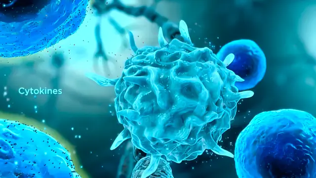
Multiplex Cytokine Assay
Ultra-Sensitive Multiplex Assay In Clinical Or Preclinical Serum/Plasma, PBMC, Tissues Etc.

Plate-Based NAb Assay
Receptor binding and functional inhibition measurement in serum/plasma

Virus Neutralization
Feasibility, Development, And Human Serum Sample Analysis

Activity/ Potency Assay
Enzymatic and binding readouts for lot characterization and release
Contact A Scientist With Your ELISA Based Assay Details
Tailored ELISA quote in 2 business days.
De-Risk Your ELISA Service With PhD Scientists For PK/PD, ADA/NAb, Cytokine, Cell Based Assays, On Budget And On Time
Kick‑Start Your ELISA Testing At Our Agile Lab!
We’ll Reach Out Within 1 Business Day- Protect your ELISA Method from costly delays. Our PhDs catch matrix interference, recovery, stability problems up front, so a flawed method never stalls your therapeutic.
- Stretch your bioanalytical budget. Proven ELISA Validation Services get your method right the first time, so your spend goes to quality science, not fixing avoidable mistakes.
- Hit your timeline. Our responsive ELISA Immunoassay Services lab keeps your study on schedule, with the scientists running your assay reachable directly through minimal management layers.
20+
Years Of ELISA Services Lab, Trusted By Biotech
500+
Quality Bioanalytical Methods, Experience Behind Every Quote
Control Cost, Hit Timeline
We develop and validate de novo ELISA assays for PK/PD, ADA/NAb, cytokine, and cell-based readouts in four to six weeks. Then about two weeks to deliver your audited report with detailed ELISA Validation documentation. You can opt for rolling data, so you act on results sooner. We handle cold-chain storage and chain-of-custody from arrival to analysis. You receive a transparent, itemized scope up front, and any scope change goes through a pre-approved SOW addendum, so costs never surprise you mid-study. We follow ICH M10 and FDA guidance unless you specify otherwise. Scientific rigor, regulatory expertise, and transparent cost on every project.
Fast Quote, Quality Work
Share details about your analyte(s) and current Enzyme Linked Immunoassay to scope your quote and timeline. Just starting? Lean on us, our ELISA Testing experience helps fill in the blanks. Have a validated kit, prior assay data, or a method to transfer? Send it, transfer is faster than de novo. Otherwise, tell us your format, sandwich, competitive, or indirect ELISA. Note sourcing for your critical reagents, proprietary or commercial capture and detection antibodies, coating, blocking, and detection reagents, yours or ours to procure. Send study protocols, sample sizes, matrix, and target sensitivity. Tell us your validation rigor, fit-for-purpose or GxP.
Reviews From Sponsors of Our ELISA Assay Validation And ELISA Assay Develpoment Services

President & CSO, Biotech
We found their integrity as refreshing as readiness to provide creative scientific input and high-quality data

Co-Founder, Biotech & University PI
NorthEast BioLab tremendously supported us in reproducing our critical lab discoveries for drug metabolism

VP, Development Operations
NorthEast BioLab’s scientists deliver high-quality data on time and within budget

VP, Biomarker Development
NorthEast BioLab is a responsive, collaborative, and reliable partner

Sr. Dir., Bioanalytical Development & QC
NorthEast BioLab offers a science-based, hands-on approach to the latest bioanalytical platforms

Executive Director, Pharmacokinetics
We trust NorthEast BioLab to design and execute streamlined, impactful bioanalytical projects
Unlock Precision, ROI With Our ELISA Service Expertise!
We’ll Respond Within 1 Business Day
Efficiently Accelerate Enzyme Linked Immunoassay For Your PK, Immunogenicity, and Multiplexed ELISA Cytokine Analysis
Expert GLP ELISA Development & ELISA Validation Services
- End-to-End Immunoassay Development: Three decades of GLP ELISA assay expertise to improve every critical parameter—antibody pairing, plate coating, signal amplification
- Up-Front Technical & Regulatory Review: We dissect your current ELISA methods, data sets, and submission needs, then deliver a written SOW and timeline within 48 hours
- In-House Reagent Tagging for Agility: Custom biotin, HRP, or fluorophore conjugation on-site boosts lot-to-lot consistency, giving you tighter CVs and wider dynamic range
Responsive ELISA Assay CRO with Tailored Lab Testing Services
- Quick-Turn Flex Scheduling: Accelerated starts and adjustable timelines accommodate urgent ELISA PK, ADA, or cytokine studies without compromising quality
- Direct Access to Senior Scientists: Real-time conversations for ELISA troubleshooting and method refinements keep potential issues from ever reaching the data table
- Dual-Layer Quality Assurance: Redundant QC and comprehensive audit trails deliver robust, reproducible ELISA results regulators trust—every single run

ELISA Assay Development & Method Development: A Step-by-Step Guide
ELISA is a powerful tool for detecting and quantifying target analytes in complex biological samples. The primary principle of ELISA assay focuses on capturing or immobilizing the target antigen on…
Our Streamlined Process For On-Time Delivery Of Entire Scope Of Work Within Sponsor Budget


Why Choose Us As Your ELISA Testing Services Lab?
FDA Audited ELISA Assay Validation, ELISA Assay Development, Or GLP Sample Analysis Bioanalytical Lab!
Partner With Our ELISA Lab For Swift, Valuable Results!
We’ll Get Back to You Within 1 Business Day- FDA-Compliant ELISA Method Development And Validation For Ligand Binding And Cell-Based Assays.
- GLP And Research ELISA Services For PK, Cytokine, And Immunogenicity Assays.
- Preclinical And Clinical ELISA Analysis For PD Biomarkers, Potency, And Functional Bioassays.
- Customized ELISA Services For Viral And Antibody Titers In Various Biological Samples.
Regulated And Inspected By:


Support Your Drug Innovation With Entire Suite OF BioAnalytical Services In Safe Hands Of Our Veteran Scientists
We Judiciously Invest In Our People, Solutions And Infrastructure, And Regularly Review Our Business Processes And Practices To Exceed Sponsor Expectations.

Related FAQs
Answers to additional ELISA Lab Services questions popular among our potential clients.