






VP, Bioanalytical Development
Services
Platforms
Solutions
About Us
 About Us
 Overview
 Leadership
 Facility & Equipment
 Quality & Compliance
 Therapeutic Areas
 Why Us?
 Values
 Process
 Partners
 Careers
Sr. Scientist – Cell Based Assay
Associate Scientist, Central Laboratory Services
Business Development Associate
Sr. Scientist, LC-MS/MS Bioanalysis – Hamden, CT, USA
Sr. Scientist, ELISA Immunoassay Bioanalysis – Hamden, CT, USA
GLP Quality Assurance Manager
Sr. Scientist - Pharmacokinetics (PK)
Content Manager, Scientific and Regulatory Writing
Resources
Interferon-alpha 2
- Interferon-alpha 2/ IFNα 2 is a cytokine secreted in response to viral infection
- IFNα 2 stimulates cellular gene expression that prevents viral spread and replication.
- IFNα 2 is a biomarker of immune and oncological disorders
- IFNα 2 can be assayed using immune assays ELISA, MSD or Luminex
Regulated And Inspected By:




Provide Your Bioanalysis Study Details
We Will Respond Within 1 Business Day
Click below for answers to your IFNα 2 Biomarker Study questions -
What is IFNα 2?
An introduction to Interferon-alpha 2 (IFNα 2)
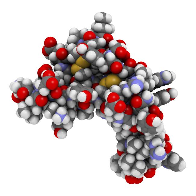
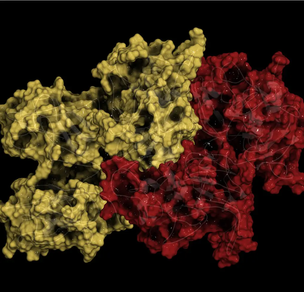
Interferon-alpha 2 or IFNα 2 is a human cytokine encoded by the IFNA2 gene. IFNA2 gene is present on chromosome 9. IFNα 2 belongs to the type I IFN family. Cells with viral infection secrete IFNα 2 and help other cells inhibit viral attack. In 1957, Alick Isaacs and Jean Lindenmann first described IFNs as a viral replication interfering agent. Type I IFN family has 13 α subtypes, out of which IFNα 2 was the very first subtypes scientists had successfully characterized in the 1980s. Therefore, researchers have widely studied IFNα 2 and the alpha interferon function to understand type I IFN family structure and mechanisms. As a result, the pharmaceutical industry also produced IFNα 2 as the first IFN drug, making it a well-known type I IFN family subtype.
The biomarker goes by other names – FNA2,IFNA, interferon, alpha 2, IFNA2B, INFA2, IFN-alphaA, IFN-alpha-2, and leIF A.
Sponsors Love Our Bioanalytical Laboratory Services For Top Quality And Industry Leading Turnaround
Connect With Us For A Reference Call With Them!

NorthEast BioLab is a responsive, collaborative, and reliable partner

We trust NorthEast BioLab to design and execute streamlined, impactful bioanalytical projects

We found their integrity as refreshing as readiness to provide creative scientific input and high-quality data

We have worked with NorthEast BioLab for over ten years given their commitment to highest quality bioanalytical data.

NorthEast BioLab tremendously supported us in reproducing our critical lab discoveries for drug metabolism

NorthEast BioLab always exceeds expectations on bioanalytical assay development, validation, and sample analysis.

“NorthEast BioLab goes the extra mile. We look forward to more meaningful collaborations.”

NorthEast BioLab offers a science-based, hands-on approach to the latest bioanalytical platforms

We are thrilled to complete our bioanalytical studies with their top quality and incredibly responsive team.

We worked closely to implement the most efficient and cost-effective bioanalytical assay for our PK Studies.

Our projects with NorthEast BioLab include successful method development, validation, stability studies during Clinical Phase I – IV studies.

NorthEast BioLab provides critical insight, and are compliant with regulatory standards and industry best practices. We highly recommend them and look forward to working together again.

Our latest successful study was a pivotal bioequivalence study, where samples from a cross-over study with about 100 volunteers needed swift analysis.

This study, same as all other bioanalytical studies, was completed with top quality and reporting standard with incredible responsiveness.

NorthEast BioLab’s scientists deliver high-quality data on time and within budget
Tackle Your IFNα 2 Biomarker Assay Testing With Our Veteran Team Now!
How do we perform IFNα2 lab test?
Protocols we follow to perform IFNα2 tests
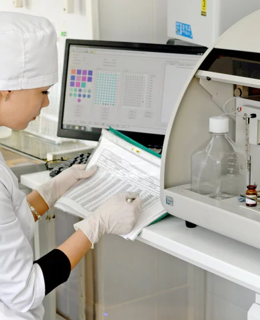
At NorthEast BioLabs, we perform Luminex, ELISA, and MSD assays to detect alpha interferons and beta interferons in plasma, serum, cell culture supernatants, tissue homogenates, cell lysates, and other fluids. Following, we have outlined a double-antibody Sandwich ELISA assay protocol.
We begin the IFN Alpha testing with a pre-coated microtiter plate with an IFNα 2 specific antibody. We add 100 μl of sample or standard to the wells and introduce a biotin-conjugated antibody in each well. This antibody is specific to IFNα 2. Next, we incubate Avidin conjugated Horseradish Peroxidase in each well. Post-incubation, we add a TMB reporter molecule. Wells containing all three, IFNα 2, enzyme-conjugated Avidin, and Biotin-conjugated antibodies will show a color change. To end the reaction we add sulphuric acid and measure the color change spectrophotometrically at 450nm (± 10nm). We then correlate optical density (O.D) of the sample, with the standard curve and determine the concentration of IFNα 2 in the samples.
The anti-interferon assay employs the principle of immunosorbent assay and colorimetric system to identify IFNα 2 in the samples. The assay has a detection range between 15.6 pg/ml – 1000 pg/ml, with minimum detectable IFNα 2 doses of typically < 6.2 pg/ml. The assay is highly precise, with an intra-assay precision of < 10% and inter-assay precision of <12%.
Registered And Inspected By Various Regulatory Agencies






Don’t Wait, Start Your IFNα 2 Biomarker Assay Testing Today!
Where could I learn more about IFNα 2 Assay ?
Popular IFNα 2 resources for you to explore
The excretion IFNα 2 from the body is rapid, and they have a brief life in the biological organism Digestive enzymes efficiently degrade oral doses of IFNα 2 effectively. Therefore, researchers predominately study the intravenous administration of the alpha interferon to determine its optimal function.
IFNα2a and IFNα2b are IFNα 2 variants, popularly known by their commercial trade names Roferon-A and Intron-A, respectively. IFNα2a and IFNα2b have shown efficiency against viral infections, some kinds of cancers, and blood disorders. As cells naturally produce IFN Alpha 2 in response to an antigen intrusion, IFNα 2 therapy also induces similar side effects in the body. Therefore, researchers progressively replace IFNα 2 treatment with well-researched drugs.
To learn more about the promising benefits of IFNα 2, explore the resources below.
http://chemocare.com/chemotherapy/drug-info/interferon-alfa.aspx
https://www.sciencedirect.com/topics/pharmacology-toxicology-and-pharmaceutical-science/alpha-interferon
https://www.macmillan.org.uk/cancer-information-and-support/treatments-and-drugs/interferon-alpha
Multiplex Cytokine Analysis, MSD Cytokine Assay
Understand Multiplex Cytokine Analysis, Meso Scale Discovery (MSD) Cytokine Assay For Inflammation, Chemokines, Interleukins, Growth Factor …

What are IFNα 2 Functions and Effects?
Understanding the functions and effects of IFNα 2
Once a virus infects a cell, the cell releases IFN alpha 2 that acts locally and on cells expressing IFNAR cell surface receptors. IFNAR comprises IFNAR1 and IFNAR2 subunits that are expressed on the surfaces of all cells. Post-receptor binding, interferon alpha 2 beta activates several cellular processes that carry the signal from the cell surface to the nucleus. This signal forms the IFN-Stimulated Gene Factor 3 complex in the nucleus, which starts the transcription of IFN-stimulated genes (ISGs).
ISGs are the core component that encodes cellular modulating proteins. ISGs have a wide range of cellular functions. They inhibit viral spread and replication, and protect uninfected cells by blocking viral entry into them. Research has discovered hundreds of interferon alpha cytokine or ISGs, explaining why type I IFN family has such a broad spectrum of biological activity. Besides, IFNα 2 inhibits cell proliferation, regulates immune system activation, and shows potent antitumor characteristics.
Trust Our IFNα 2 Biomarker Assay Testing.
Explore More Biomarkers
-
Brain-Derived Neurotrophic Factor
-
 Granulate Colony Stimulation Factor
-
 Epidermal growth factor
-
 Fas Ligand
-
 Granulocyte Macrophage Colony Stimulating Factor
-
 Leukemia Inhibitory Factor
-
 Luteinizing hormone
-
 Macrophage Migration Inhibitory Factor
-
 Interferon Beta
-
 Interferon Gamma
-
 Transforming Growth Factor Beta 1
-
 B Type Natriuretic Peptide
-
 Glial Fibrillary Acidic Protein
-
 Cystatin C
-
 Hepatocyte Growth Factor
-
 Interferon Alpha 2
-
 Tumor Necrosis Factor Alpha